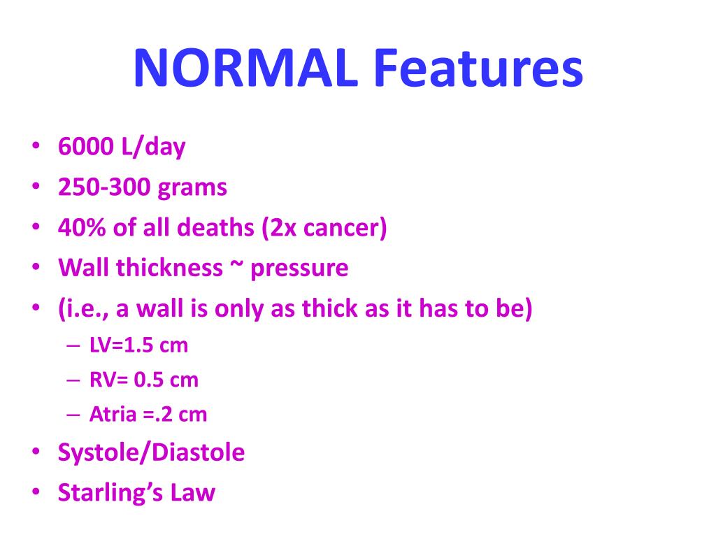
Normal Rv Wall Thickness

Normal Values For The Right Ventricle Rv Linear And Area Dimensions 18 Download Table

Table 2 From Anatomy Echocardiography And Normal Right Ventricular Dimensions Semantic Scholar
Echo Assesment Of Rv Function

Simulation Results Ventricular Wall Thickness Download Table

Body And Heart Weights And Ventricular Wall Thickness In Normal And Download Table
Echocardiographic Assessment Of The Right Heart In Adults Ppt Video Online Download
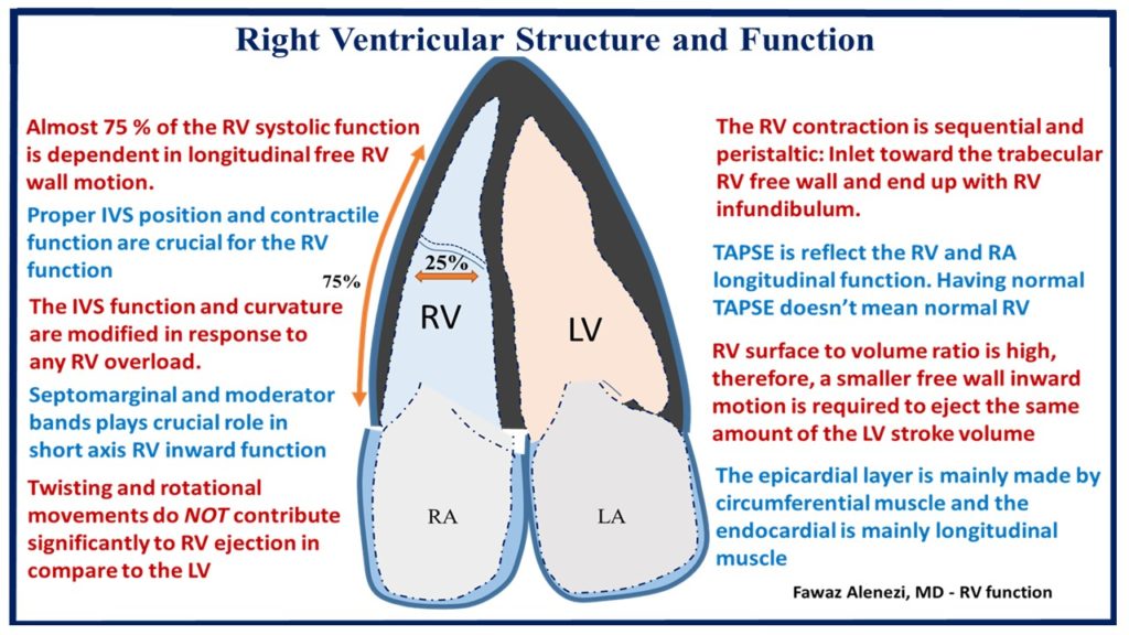
This is partially due to the rv s complex architecture which makes quantification of rv mass by echo difficult.
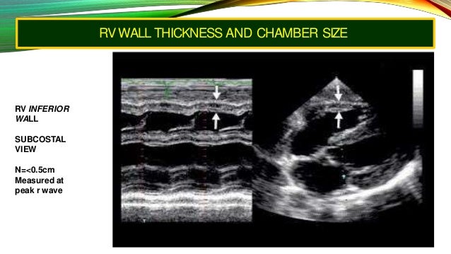
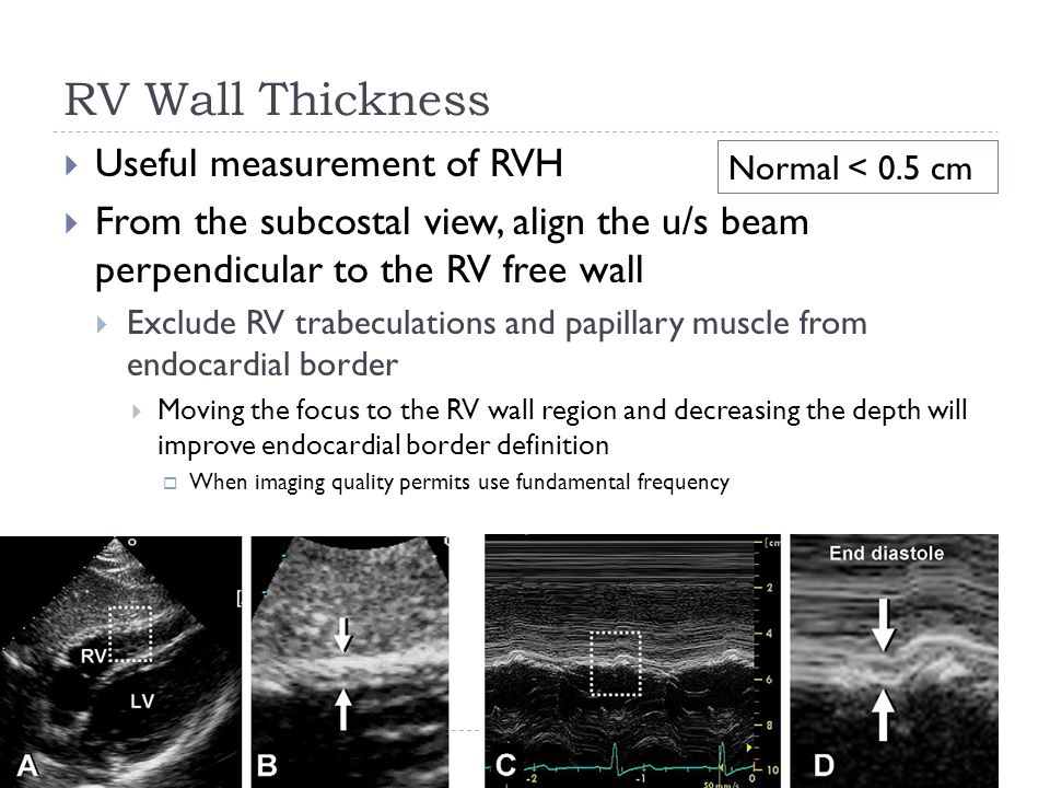
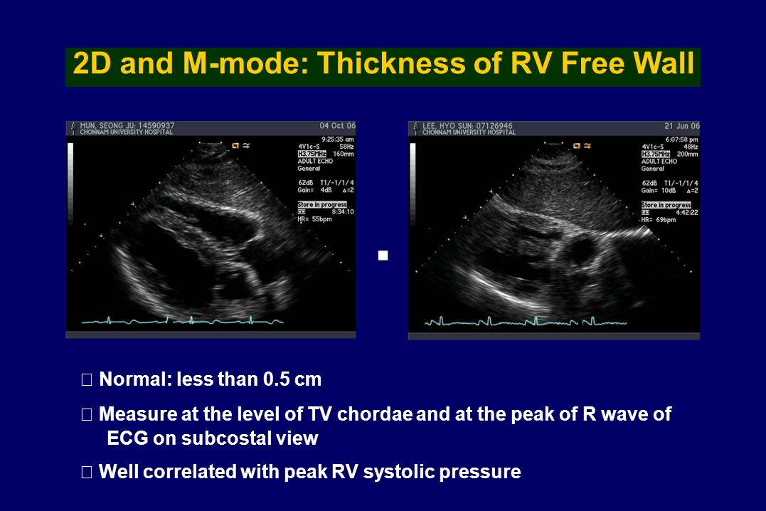
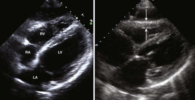
Normal rv wall thickness. Rv wall thickness is measured in diastole pref erably from the subcostal view using either m mode or two dimen sional 2d imaging figure 5. Here we retrospectively evaluate the thickness of the inferior rv wall irvwt by echo in neonates and infants with normal. An established echocardiographic echo standard for assessing the newborn right ventricle rv for hypertrophy has not been thoroughly developed. Normal is less than 5 mm best is ps la second is subcostal.
Rv medio lateral end diastolic dimension 4 3 cm rv end diastolic area 35 5 cm 2 maximal ra medio lateral and supero inferior dimensions 4 6 cm and 4 9 cm respectively maximal ra volume 33 ml m 2 35 89. The feasibility of subxiphoid echocardiography to measure the thickness of the right ventricular wall rvwt was investigated. Rv medio lateral end diastolic dimension 4 3 cm rv end diastolic area 35 5 cm 2 maximal ra medio lateral and supero inferior dimensions 4 6 cm and 4 9 cm respectively maximal ra volume 33 ml m 2 35 89.
Normal 2d measurements from the apical 4 chamber view. Echo assessment of rv 3 0 2 5 3 8. How does acute rv enlargement differ from chronic rv enlargement. Rv and or lv dysfunction using the normal cut off of.
Normal range on the basis of a right ventricle appearing significantly larger than the left ventricle. Here is a five star rated article on rv dimension. Moody jr md uthscsa and almmvah october 2001. Alternatively the left parasternal view is also used for measuring rv wall thickness.
Abnormal rv wall thickness should be reported in patients suspected of having. The interior wall panel wood or aluminium framing polystyrene insulation and exterior wall material which can be filon a plastic and fibreglass combined sheeting will all be affixed with a strong industrial adhesive and fed through a pinch. Normal or increased thickness is expected in chronic rv enlargement. In 87 90 6 of the 96 patients studied adequate visualization of the echoes from the right ventricular wall was obtained using the subxiphoid technique.
Echo assessment of rv walls weyman 1994 p. Assessing rv thickness size and function joe m. Normal 2d measurements from the apical 4 chamber view. Thickness 5 mm indicates rv hypertrophy rvh and may suggest rv.
Dilatation is more conspicuous in acute rve pulmonary embolism rv infarct associated wall motion defects and thinning favors acute rve.
Assessment Of The Right Ventricle By Echocardiography Ppt Download

Right Heart Thoracic Key
Echocardiography In The Patient With Right Heart Failure Thoracic Key
Left Ventricular Systolic Performance And Pathology Radiology Key
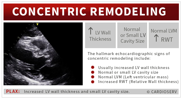
Understanding Lvh Part 1 Concentric Eccentric And Concentric Remodeling
Https Www Ahajournals Org Doi Pdf 10 1161 01 Cir 56 2 278

Relative Wall Thickness And The Risk For Ventricular Tachyarrhythmias In Patients With Left Ventricular Dysfunction Jacc Journal Of The American College Of Cardiology

Right Ventricle Right Atrium Tricuspid And Pulmonic Valves Anesthesia Key
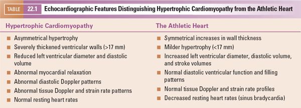
Hypertrophic Cardiomyopathy Thoracic Key

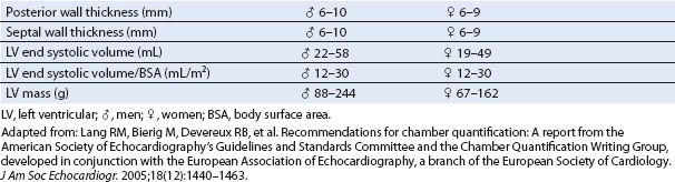
S Systole D Diastole Lvd Left Ventricle Dimension St Septal Thickness Pwt Posterior Wall Cardiac Sonography Ultrasound Technician Cardiac Anatomy

Normal Reference Values Of Echoca Preview Related Info Mendeley
Reference Normal Values For Echocardiography Ecg Echo

Right Ventricular Hypertrophy Wikipedia

Left Ventricle An Overview Sciencedirect Topics
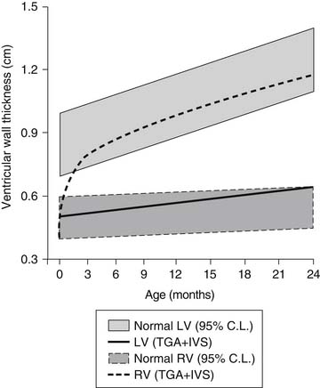
Transposition Of The Great Arteries Simple And Complex Forms Thoracic Key

Pdf Normal Values Of M Mode Echocardiographic Measurements Of More Than 2000 Healthy Infants And Children In Central Europe

Deposit Diseases As Differential Diagnosis Of Left Ventricular Hypertrophy In Patients With Heart Failure And Preserved Systolic Function

Sports Cardiology Core Curriculum For Providing Cardiovascular Care To Competitive Athletes And Highly Active People Sciencedirect
Https Encrypted Tbn0 Gstatic Com Images Q Tbn 3aand9gcs2zqlvslkgulmpnrjjpswwwcc6oodz66mffjk5yypss3w Njur Usqp Cau
Acute Right Heart Failure Adaptation Interdependence And External I

Left Ventricle Lv Size And Function Pdf Free Download
Https Onlinelibrary Wiley Com Doi Pdf 10 1002 Jum 14516
Lvef The Early Career Voice

Role Of Right Ventricular Wall Motion Abnormalities In Risk Stratification And Prognosis Of Patients Referred For Stress Echocardiography Jacc Journal Of The American College Of Cardiology
Https Www Ahajournals Org Doi Pdf 10 1161 01 Cir 60 5 1058
Https Www Ajronline Org Doi Pdf 10 2214 Ajr 12 9334

Acvim Consensus Statement Guidelines For The Classification Diagnosis And Management Of Cardiomyopathies In Cats Luis Fuentes 2020 Journal Of Veterinary Internal Medicine Wiley Online Library
Https Onlinelibrary Wiley Com Doi Pdf 10 1111 Echo 13491

Heart Basicmedical Key

Spectrum Of Restrictive And Infiltrative Cardiomyopathies Part 1 Of A 2 Part Series Sciencedirect

Right Ventricle An Overview Sciencedirect Topics
Https Www Ahajournals Org Doi Pdf 10 1161 01 Res 20 6 649
Https Www Ahajournals Org Doi Pdf 10 1161 01 Cir 61 2 441 Download True
Left Ventricular Geometric Patterns In Hypertensive Nigerians A Systematic Review Karaye International Cardiovascular Forum Journal

Right Ventricular Hypertrophy Is An Increase In The Thickness Of Chronic Obstructive Pulmonary Chronic Obstructive Pulmonary Disease Congenital Heart Disease
Inherited Cardiomyopathies And Sports Participation Springerlink

Maternal Nutrient Restriction During Pregnancy And Lactation Leads To Impaired Right Ventricular Function In Young Adult Baboons Kuo 2017 The Journal Of Physiology Wiley Online Library

Afterload An Overview Sciencedirect Topics

Hypertrophic Cardiomyopathy 2016 Update
Https Www Onlinejase Com Article S0894 7317 16 30677 0 Pdf

Guidelines And Standards Pdf Free Download
Https Journals Sagepub Com Doi Pdf 10 1177 1753465815621251